CASEⅠ. Cleanlant implant 11.4 years F/U in the mandibular posterior region
Fig. 1.
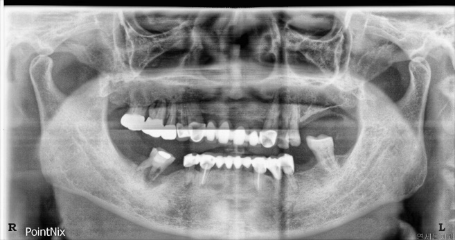
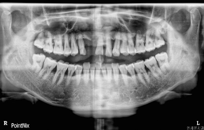
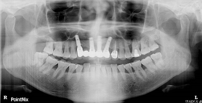
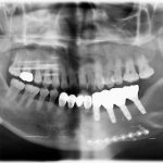
Pre-op panorama. A 37 years male patient visited clinic for fractured root rest extraction and implant surgery.
CASEⅠ. Cleanlant implant 11.4 years F/U in the mandibular posterior region
Fig. 2.
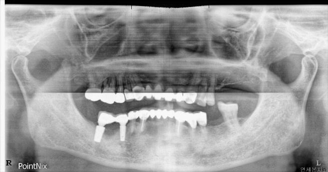
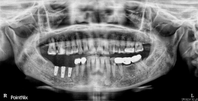
Post-op panorama(Mar. 13, 2007). #46 Extraction, and immediate implant placement with buccal and vertical bone augmentation. (DENTIS Cleanlant Ø4.3 x 10mm on #45, Ø6.0 x 10mm on #47)
CASEⅠ. Cleanlant implant 11.4 years F/U in the mandibular posterior region
Fig. 3.
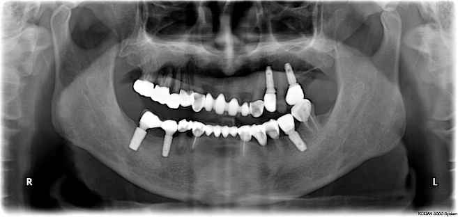
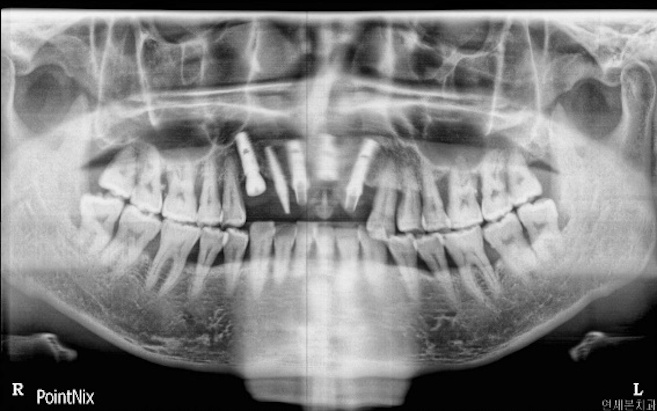
4 years implant f/u panorama(Jan. 25, 2011). There is no significant sign. The peri-implant bone level is similar to before.
CASEⅠ. Cleanlant implant 11.4 years F/U in the mandibular posterior region
Fig. 4.
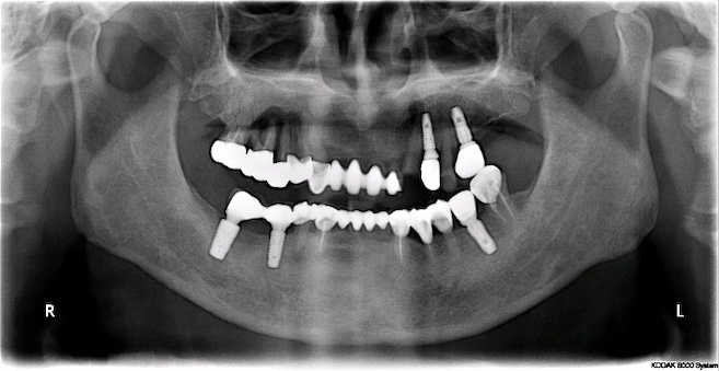
8.5 years implant f/u panorama(Oct. 01, 2015). There are no abnormal findings such as bone resorption. The bone at the defect area was regenerated.
CASEⅠ. Cleanlant implant 11.4 years F/U in the mandibular posterior region
Fig. 5.
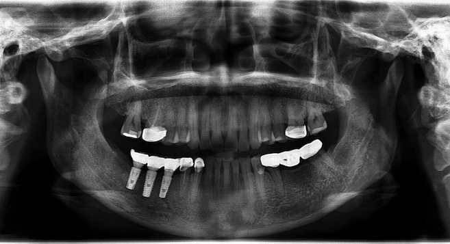
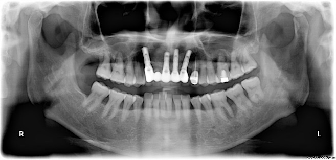
11.4 years implant f/u panorama(AUG, 2018). There are no abnormal findings such as bone resorption. The peri-implant bone level remains stable, and it is a normal state.
CASEⅡ. Cleanlant implant 7.6 years F/U in the mandibular posterior region
Fig. 6.
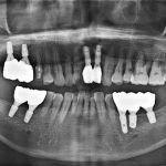
Post-op panorama(Mar. 12, 2009). A 53 years female patient visited for implant placement and buccal bone augmentation. (DENTIS Cleanlant Ø4.5 x 10mm on #45, Ø5.0 x 12mm on #46, Ø5.0 x 10mm on #47)
CASEⅡ. Cleanlant implant 7.6 years F/U in the mandibular posterior region
Fig. 7.
7.6 years implant f/u panorama (Oct. 26, 2016). There are no abnormal findings. The peri-implant bone level remains stable.
CASEⅢ. Cleanlant implant 6 years F/U in the maxillary anterior region
Fig. 8.
Pre-op panorama. A 40 years male patient visited for implant surgery in maxillary anterior.
CASEⅢ. Cleanlant implant 6 years F/U in the maxillary anterior region
Fig. 9.
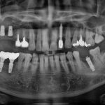
Post-op panorama(Aug. 24, 2010). Implant placement with DENTIS Cleanlant Ø4.3 x 10mm on #11, #13, #21, #22, and DENTIS I-FIX Angled type on #12 for temporary crown. And #13, #21 bone augmentation.
CASEⅢ. Cleanlant implant 6 years F/U in the maxillary anterior region
Fig. 10.
1 year implant f/u panorama(Sep. 02, 2011). There are no abnormal findings such as bone resorption. The peri-implant bone level remains stable.
CASEⅢ. Cleanlant implant 6 years F/U in the maxillary anterior region
Fig. 11.
6 years implant f/u panorama (August 23, 2016). There are no abnormal findings. The peri-implant bone level remains stable, and it is a normal state.
The copyright of this clinical data belongs to Dr. Lee, Kwangwon in Central Yonsei Dental Clinic, Korea. Please note that the copyright of this card news and the right of use belongs to DENTIS Co., Ltd, GDIA.