Prof. Moon, Seong-yong
Chosun University Dental Hospital
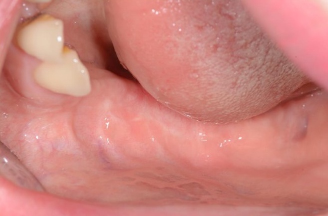
Fig. 1.
Pre-op clinical view. 41 years old female patient visited for implant consultation.
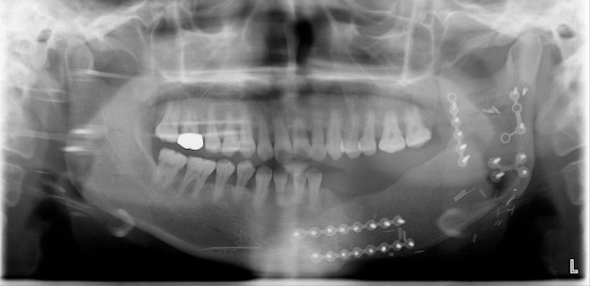
Fig. 2.
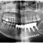
Pre-op panorama. The lower left bone and teeth were severely damaged following surgery to re-move the tumor.

Fig. 3.
Implant placement simulation and SIMPLE GUIDE was planned by BlueSkyBio Software.
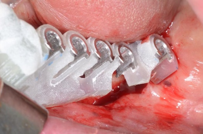
Fig. 4.
SIMPLE GUIDE was fabricated by SLA 3D printer, ZENITH U.
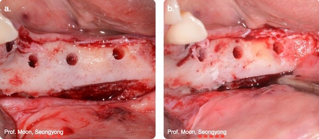
Fig. 5a-b.
Pilot drilling was done by SIMPLE GUIDE KIT(a). Final drilling was done by OneQ surgical KIT(b).
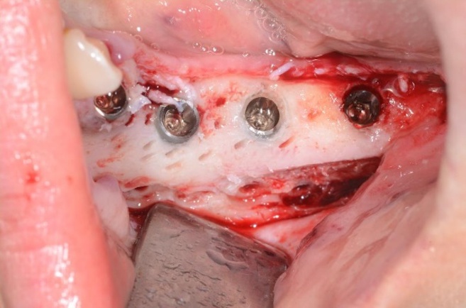
Fig. 6.
OneQ-SL implants were placed on #33, #34(Ø4.2 x 10mm), #35, #36(Ø4.7x10mm).
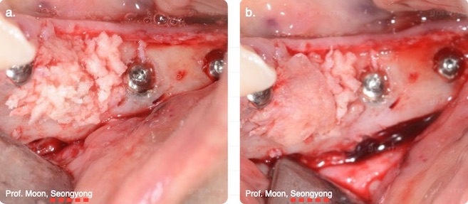
Fig. 7a-b.
The autogenous bone graft(a) and PRF application(b).
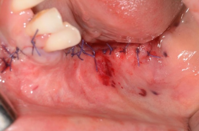
Fig. 8.
Suture was done.
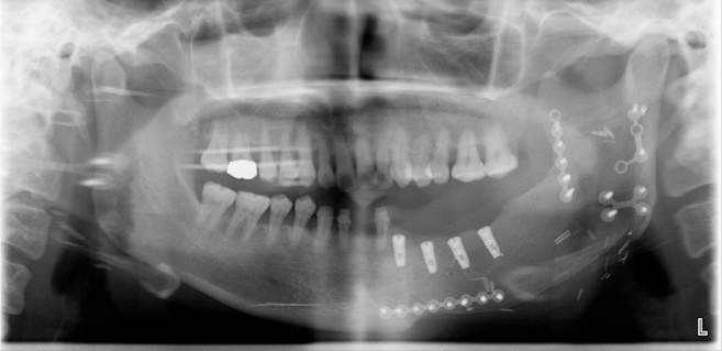
Fig. 9.
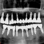
Post-op panorama.
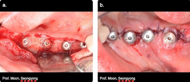
Fig. 10a-b.
Post-op 4 months. 2nd surgery was done(a). Healing Abutments and Louis ButtonⅡ were connected(b).
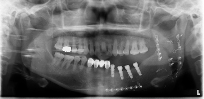
Fig. 11.
Post-op 4 months panorama.
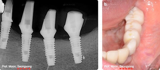
Fig. 12a-b.
Post-op 10 months. Customized abutments(a) and provisional prosthesis(b) were delivered.
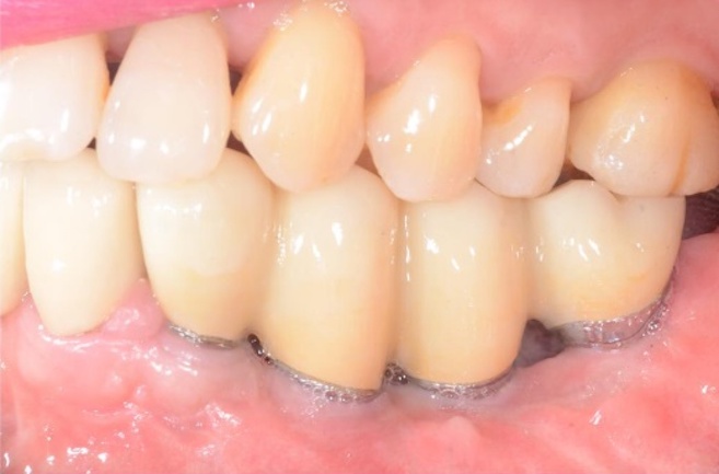
Fig. 13.
1 year after implant placement, Final prosthesis was delivered.
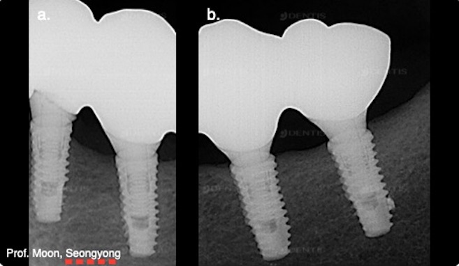
Fig. 14a-b.
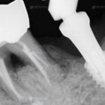
Radiography of #33, #34(a), #35, #36(b) Final prosthesis.
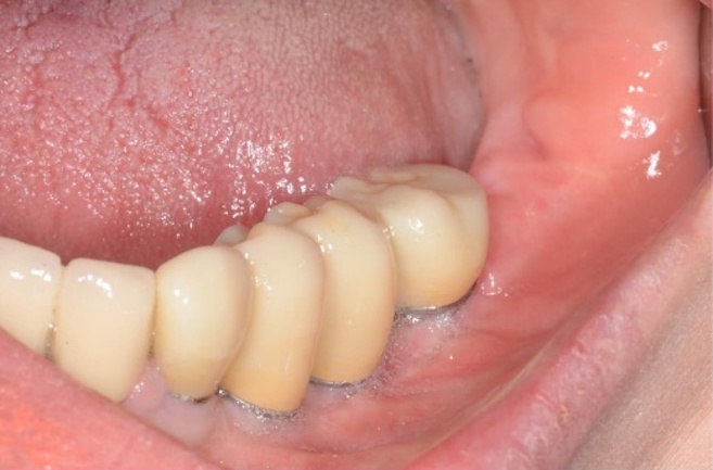
Fig. 15.
Post-op 1.6 years clinical view.
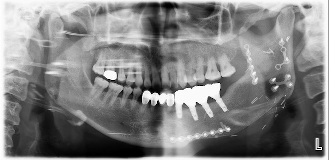
Fig. 16.
Post-op 1.6 years panorama was normal.
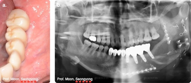
Fig. 17a-b.
Post-op 2.4 years clinical(a) and panoramic(b) view. There was no abnormal sign.
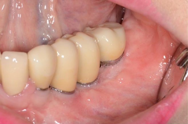
Fig. 18.
Post-op 2.9 years clinical view.
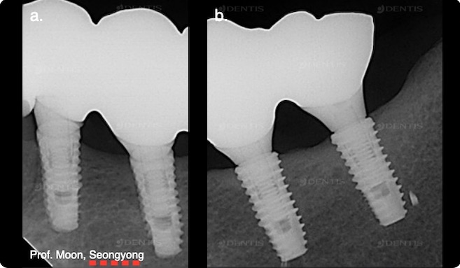
Fig. 19a-b.
Post-op 2.9 years radiography of #33, #34(a), #35, #36(b). There was no change in bone around fixtures.
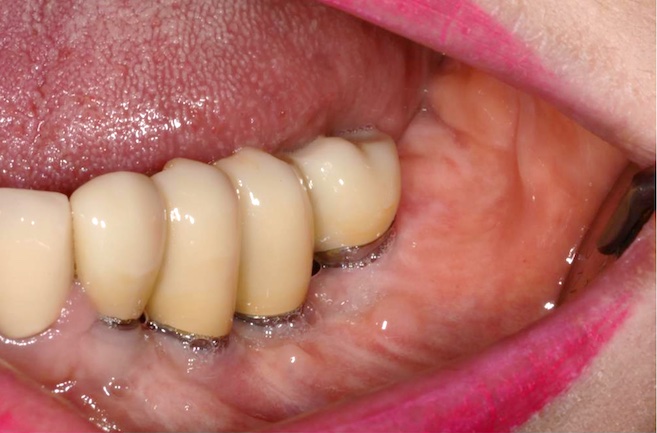
Fig. 20.
Post-op 3.4 years clinical view.
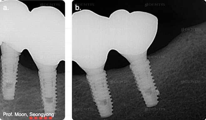
Fig. 21a-b.
Post-op 3.4 years periapical X-ray of #33, #34(a), #35, #36(b). There was no significant abnormality.
The copyright of this clinical data belongs to
Prof. Moon, Seongyong in Chosun University Dental Hospital.
Please note that the copyright of this card news
and the right of use belongs to DENTIS Co., Ltd and GDIA.
